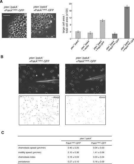
FIGURE 6:
Phosphorylation of PakA at its PKB substrate motif site is required for its function. PakAT585A-GFP or PakAT585E-GFP was expressed in pten-/pakA- cells. (A) Representative images of cells on glass substrates. Relative area occupied by multinucleated cells are shown (n > 55). Gray bars are replotted from Figure 5A. (B) Chemotaxis assays. Cells were observed for 45 min at 30-s intervals. Upper panels show images at 45 min after chemotaxis was initiated. Lower panels show trajectories of the entire recording field. Bar, 50 μm. (C) Quantification of the chemotaxis movies. Average and standard deviation of at least three movies for each cell line are calculated. See Material and Methods for the definitions of these parameters.

