FIGURE 5:
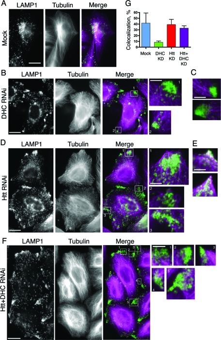
Htt may play a role in coordinating organelle association with microtubules. HeLa cells were mock transfected (A) or depleted of DHC (B) and stained for LE/lysosomes (LAMP1) and microtubules. The magnified boxed regions (boxes 1 and 2) show that the peripheral LE/lysosomes are most often found just beyond the tips of the microtubules at the cell periphery. (C) Additional merged images (as in B) featuring magnified regions of peripheral LE/lysosomes and microtubules. (D) HeLa cells depleted of Htt were stained as in (A). The magnified boxed regions (boxes 1–3) show that the peripheral LE/lysosomes are most often found enmeshed in regions dense with microtubules. (E) Additional merged images (as in D) featuring magnified regions of overlapping LE/lysosomes and microtubules at the cell edge. (F) HeLa cells depleted of Htt and dynein were stained as in (A). The magnified boxed regions (boxes 1–4) show that the peripheral LE/lysosomes are most often found enmeshed in regions dense with microtubules, similar to (D). Scale bar = 20 μm, except magnified images. The magnified images are 4× the size of original image; scale bar = 5 μm. (G) Colocalization of peripheral patches of LE/lysosomes and microtubules was quantified in mock (n = 4), DHC RNAi (n = 17), Htt RNAi (n = 4), and Htt and DHC double-knockdown cells (n = 16). Error bars, ±SEM.

