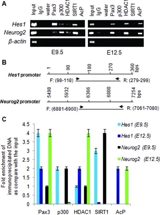
FIGURE 5:
SIRT1 binds to Hes1 and Neurog2 promoters from E9.5, but not E12.5, mouse caudal neural tube. (A) ChIP assays were done with E9.5 and E12.5 mouse lumbar neural tube. ChIP compatible antibodies against Pax3, p300, HDAC1, SIRT1, and acetylated protein (AcP) were used to immunoprecipitate (IP) the protein–DNA complex. IgG was used as an IP-negative control. Murine β-actin primers were used as negative loading control. Amplified product was present only in the input and not in the control IgG or the immunoprecipitate. (B) The 200-bp amplified products using Hes1 and Neurog2 promoter primer sets are shown. Each ChIP experiment was performed in triplicate using one lumbar neural tube region per ChIP assay with a total of n = 4. (C) Immunoprecipitated DNA was subjected to quantitative PCR using murine primers for Hes1 and Neurog2 promoters. The data represent fold enrichment of immunoprecipitated DNA compared with input sample. Each ChIP experiment was performed in triplicate using one lumbar neural tube region per ChIP assay with a total n = 4.

