FIGURE 3:
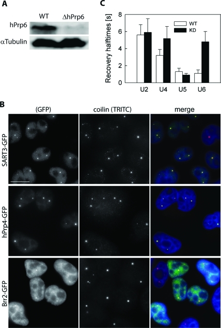
hPrp6 KD. (A) HeLa cells were treated with siRNA specific for hPrp6 mRNA, and the level of hPrp6 protein was determined by Western blotting before and after siRNA KD. α-Tubulin served as a loading control. (B) Depletion of hPrp6 resulted in accumulation of SART3-GFP and hPrp4-GFP in CBs (depicted by coilin immunostaining). Localization of Brr2-GFP was not significantly altered. Compare with Figure 1A. Bar: 10 μm. (C) Mobility of GFP-tagged snRNP markers was monitored in CBs by FRAP, and mean fluorescence recovery halftimes (t1/2) were calculated in untreated cells (empty bars) and after hPrp6 KD (full bars). The halftime values were obtained from double-exponential fits of the measured fluorescence intensities. Mean values and standard deviations (error bars) were calculated from at least 10 FRAP curves for each cell line.

