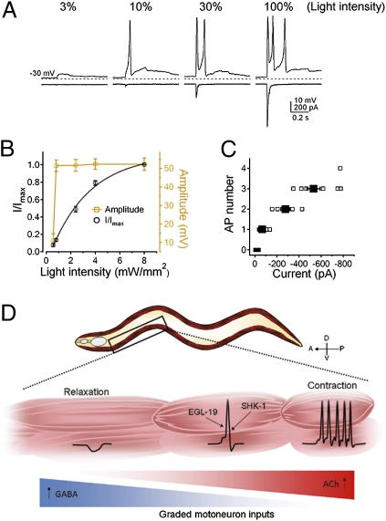
Fig. 6.
Action potential-driven muscle contraction and relaxation in response to graded motoneuron inputs. (A) Graded postsynaptic currents (at −30 mV) evoked all-or-none action potentials (at 0 pA). 3%, 10%, 30%, and 100% indicate the percentage of the full light stimulation. Dashed lines, −30 mV. Data were recorded from the same muscle cell every 30 s. (B) Normalized postsynaptic currents (○, n = 14) and corresponding membrane potential peak amplitude (□, n = 11) were plotted against light intensity. The normalized postsynaptic currents were fitted with a single exponential function. (C) The number of action potentials plotted against the amplitude of postsynaptic currents (n = 8). (Error bars = SEM.) (D) Graphical representation of a model: In response to graded motoneuron inputs, muscle cells fire action potentials that coordinate the contraction or relaxation along the body.

