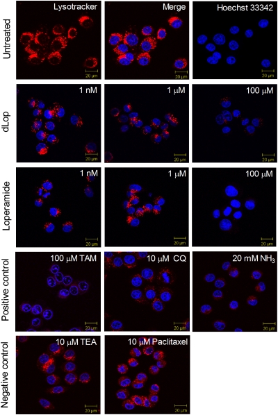
Fig. 2.
Reduction in accumulation of a fluorescent weak base from lysosomes by increasing concentrations of dLop and loperamide as assessed by confocal microscopy. Images are merged to show the simultaneous staining of the lysosomes with LysoTracker Red DND-99 (10 nM) and of the nucleus with Hoechst 33342 (8 μM; row 1). Weak bases dLop, loperamide, tamoxifen (TAM), chloroquine (CQ), and ammonia (NH3) block the accumulation of the fluorescent base (rows 2–4); the cationic compound TEA-HCl and the nonbase paclitaxel do not (row 5).

