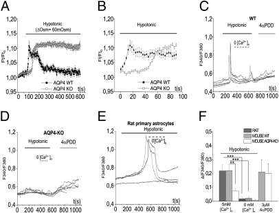
Fig. 1.
AQP4 is essential for RVD and for the hypotonicity-induced [Ca2+]i response in primary astroglial cells in primary astroglial cells. (A) Calcein-quenching method for measurement of osmotically induced volume changes in primary astroglial cells from WT (closed symbols, n = 16) and AQP4-KO mice (open symbols, n = 30). The cells were exposed to a hypotonic medium (ΔOsm = 60 mOsm). (B) The initial parts of the curves in A, shown at higher time resolution. Zero time point on the x axis corresponds to 90 s in Fig. 1A. Quantitative analyses of data in A and B are shown in Fig. S1. (C–E) Typical [Ca2+]i dynamics recorded in fura-2–loaded primary astroglial cells from WT mouse (C), AQP4-KO mouse (D), and rat (E). Each line represents the response of an individual cell. (F) Quantitative analyses of the hypotonicity-induced [Ca2+]i response in primary astrocytes. Rat astrocytes (n = 15) are shown by the dark gray bar, WT mouse astrocytes (n = 17) by the light gray bar, and AQP4-KO astrocytes (n = 14) by the white bar. P = 0.0016 for the comparison between AQP4 WT and AQP4-KO astrocytes, independent t-test; P < 0.001 for experiments in presence and absence of [Ca2+]o, paired t test. The 4αPDD response did not differ significantly between WT and AQP4-KO cells (P = 0.9, independent t-test). **P ≤ 0.01; ***P ≤ 0.001.

