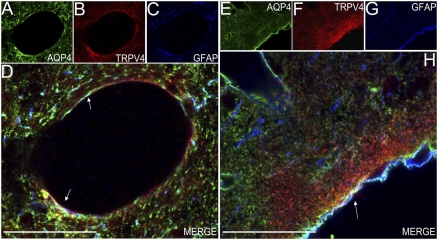
Fig. 3.
TRPV4 and AQP4 colocalize in astrocytes of adult rat brain. (A–C) Single-plane confocal immunofluorescence images of large-caliber blood vessel of rat occipital cortex. Triple labeling with goat anti-AQP4 (green, A), rabbit anti-TRPV4 (red, B), and mouse anti-GFAP (blue, C) reveals astrocyte processes that are immunopositive for TRPV4 and AQP4. Triple-labeled processes appear in white in D. (Scale bar: 50 μm.) (E–G) Single-plane confocal immunofluorescence images of sagittal section of rat occipital cortex. Triple labeling with goat anti-AQP4 (green, E), rabbit anti-TRPV4 (red, F), and mouse anti-GFAP (blue, G). Triple-labeled processes appear in white in H (merged image, arrow). The triple-labeled processes (white arrows) face the pial surface and surround large-caliber blood vessels. (Scale bars: 50 μm.)

