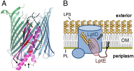
Fig. 5.
Proposed models for the structure and function of the LptD/E complex. (A) LptE forms a plug within the lumen of the LptD β-barrel. A model LptE structure (as in Fig. 2B) is shown in magenta, with the LptD-interacting residues depicted as red sticks and with an asterisk at the N-terminus of the model structure. The transmembrane portion of LptD is shown as a hypothetical 22-stranded β-barrel in blue-green, with the LptE-interacting extracellular loop as a dashed black line. Arrows denote approximate locations of the trypsin cleavage sites within LptE in LptD/E complexes (10). (B) Proposed model for direct insertion of LPS into the outer leaflet of the OM. LPS is delivered directly from the periplasm to LptE within the LptD pore, and subsequently into the OM lipid bilayer through an opening in the LptD β-barrel wall (dashed blue line), without residing in the inner leaflet. PL, phospholipid. The O-antigen of LPS has been omitted for clarity.

