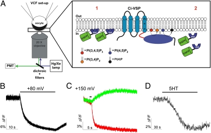
Fig. 1.
Development of cell-based assay to study cell membrane binding of GFP-fusion proteins. (A) Measuring membrane binding of GFP protein. Xenopus oocytes expressing the protein of interest are voltage clamped. The fluorescence of the EGFP-fusion protein is recorded by epifluorescence (1). The cortical layer of pigment granules in the animal hemisphere and the opaque cytoplasmic compartment provide a mask for cytoplasmic fluorescence (2). When the fusion proteins lose their interaction with the plasma membrane, the fusion proteins diffuse into the cell, inducing a decrease of fluorescence. (B–D) PH-PLC domain assay. Representative example of EFGP-PHPLC fluorescence decrease induced by Ci-VSP activation (30-s pulse from −80 to +80 mV) (D) and 5HT2cR activation (C) in Xenopus oocyte. (C) Representative example of tagRFP-PHPLC (red) fluorescence decrease and EFGP-PHOSH1 fluorescence increase (green) induced by Ci-VSP activation (200-ms pulse from −80 to +150 mV).

