Fig. 3.
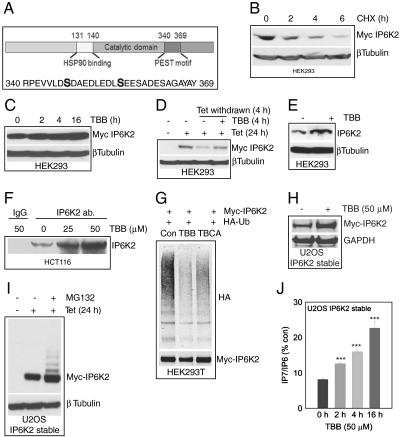
IP6K2 stability is enhanced by CK2 inhibition A. CK2 phosphorylation sites at the degradation specific PEST motif of IP6K2. B. Myc-IP6K2 protein displays a half-life of 2 h as measured by cycloheximide treatment of HEK293 cells. C. TBB (50 μM) stabilizes Myc-IP6K2 protein levels in a time-dependent fashion. D. TBB (50 μM) protects tetracycline removal induced IP6K2 protein degradation. E. TBB (50 μM, 6 h) stabilizes endogenous IP6K2 levels in HEK293 cells. F. Immunoprecipitation of endogenous IP6K2 from HCT116 cells reveals increased protein levels after TBB treatment. G. IP6K2 ubiquitination is significantly reduced after TBB (50 μM) or TBCA (10 μM) treatment, H. TBB stabilizes Myc-IP6K2 in U2OS cells. I. The MG132 treatment reveals multiple ubiquitinated Myc-IP6K2. J. TBB increases IP7 levels in U2OS cells stably expressing Myc-IP6K2. (***p < 0.001).

