Fig. 2.
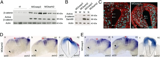
Barhl2 and Casp3 depletion increases β-catenin and Wnt activity. (A and B) Western blot analysis. Proteins (10 μg) extracted from st 26 heads of embryos injected with GFP (ct), MOcasp3, or MObarhl2. α-actin (42 kDa) is used as a loading ct. (A) Barhl2- and Casp3-depleted head extracts exhibit a significant increase in both β-catenin and active β-catenin levels (95 kDa) as indicated. (B) Active β-catenin detected in MOcasp3- or MObarhl2-injected head extracts is enriched in nuclear head extracts (N) compared with that in whole-cell head extracts (W). Sam68 is used as a marker of proper nuclear proteins extraction. (C) β-catenin mislocalization. Representative IHC images for β-catenin (red) merged with BB (blue) on diencephalic sections of st 26/27 embryos injected with MObarhl2 or MOcasp3. Arrows indicate the part of apical surfaces where β-catenin staining is disrupted. Inj, injected side. (D and E) Analysis of Wnt activity. axin2 expression profiles at st 30 analyzed by ISH. The ct (a) and injected (b) sides of one representative dissected neural tube flat-mounted or (c) a section at the diencephalic level are shown. The percent of embryos exhibiting the phenotype is indicated. (Da) ct and (Db and Dc) Barhl2-depleted (80%, n = 15) and (Ea) ct and (Eb and Ec) Casp3-depleted neural tubes (78%, n = 18). The white rectangle delineates the prosomere P2 alar plate. The black arrow indicates localization of the optic stalk.

