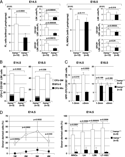
Fig. 3.
Phenotypic characterization and functional assays of hemp+/+ and hemp−/− FL cells. (A) Comparison of total cell numbers (Left) and absolute cell numbers for LSK (Right Top) CD150+ LSK (Right, Middle) and EPCR+ LSK (Right, Bottom) fractions between hemp+/+ and hemp−/− FLs at E14.5 and E18.5 (data ± SD). At E14.5, FL cells were collected without using lymphoprep gradients (Lymphoprep), whereas at E18.5, FL mononuclear cells (MNCs) were separated with the use of Lymphoprep. (B) Average numbers of colony forming units in culture (CFU-C) generated from hemp+/+ and hemp−/− CD150+ LSK cells at E14.5 and E18.5. One hundred and fifty cells were cultured for 8 d in a semi-solid culture with cytokines, and the colony types formed were classified into colony-forming unit-granulocyte, macrophage [(CFU-GM) open bar], burst-forming unit-erythoid [(BFU-E) gray bar], CFU-Mix (closed bar) and counted (data ± SD). (C) Average numbers of high proliferative potential-colony forming cells (HPP-CFCs) generated from hemp+/+ (open bar) and hemp−/− (closed bar) CD150+ LSK cells at E14.5 and E18.5. One hundred and fifty cells were cultured for 16 d in a semi-solid culture with cytokines, and colonies from 1–2 mm and >2 mm were classified and counted independently (data ± SD). (D) Competitive repopulation assay of hemp+/+ (open box) and hemp−/− (closed box) CD150+ LSK cells at E14.5. One hundred and fifty cells (Ly5.2) were transplanted into recipient mice (Ly5.1) together with 4×10⁵ competitors (Ly5.1). Donor-derived chimerism in the peripheral blood (PB) at 1, 2, 3 and 4 months after transplantation and chimerism in the MNC, Lin−, LSK and LT-HSC fractions in the bone marrow (BM) at 4 mo after transplantation are shown in the left and right panels, respectively (data ± SD).

