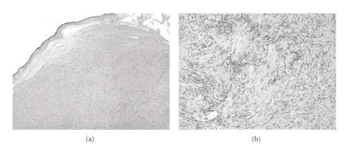
Figure 3.
Histological examination. (a) shows an encapsulated mass consisting of monomorphic spindle cells with pointed basophilic nuclei (Antoni A tissue), set in a variable collagenous stroma (low power). Given limited excision of the encapsulated mass, adjacent normal breast parenchyma is not visualized. (b) shows areas of cells with parallel arrays of nuclear palisading known as Verocay bodies (high power).

