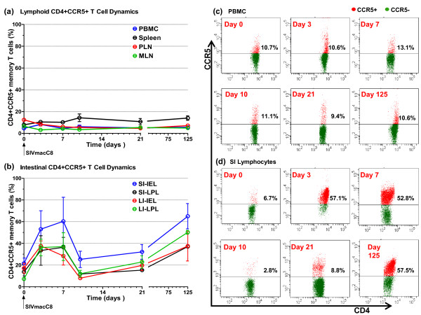
Figure 3.
CCR5+ T cell dynamics in macaques vaccinated with attenuated SIV. Following vaccination of cynomolgus macaques (n = 20) with live attenuated SIV peripheral blood, lymphoid tissue and intestinal lymphocyte CD4+CCR5+ memory T cell percentages was determined at days 0, 3, 7, 10, 21 and 125 post inoculation. There was no evidence of dynamic changes in percentages of CD4+CCR5+ memory T cell in peripheral blood and lymphoid tissues (a). In contrast, dynamic changes in CD4+CCR5+ memory T cell percentages was observed in the lamina propria and intraepithelial lymphocytes of both the small and large intestine (b). Panels (c) and (d) shows representative staining for CCR5 on CD4+ PBMC and SI lymphocytes, respectively, at each time point. CCR5+CD4+ T cells are shown in red and CCR5-CD4+ T cells in green. For analysis of peripheral blood n = 16 at day 3 reducing to n = 6 by day 21 as animals were sacrificed, n = 2 at all time points thereafter. For analysis of tissues n = 4 at all time points except days 7 and 125 where n = 2. Error bars shown are ± 1 SEM. PBMC: peripheral blood mononuclear cells, PLN: peripheral lymph node, MLN: mesenteric lymph node, SI: small intestine, LI: large intestine, IEL: intraepithelial lymphocytes, LPL: lamina propria lymphocytes.

