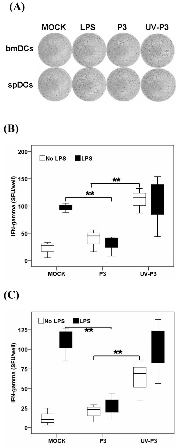
Figure 5.
IFN-γ producing T cells were detected by ELISPOT assay. P3-infected, UV-P3-stimulated or mock-treated DCs as well as differently treated spDCs were harvested and treated with Mitomycin C (Sigma-Aldrich, MO) at final concentration of 10 μg/ml for 1 h. The differently treated or mock DCs were seeded (1 × 10⁴ per well) together with 1 × 10⁵ per well T cells in triplicates for 20 h. LPS-stimulated DC/T cell co-cultures served as positive controls. One representative for IFN-γ spot forming unit (SFU) by ELISPOT assay was shown (A). The figure was representative of three independent experiments. Corrected data (SFU)/well were shown for bmDCs and spDCs activations for naïve T cells to expand and produce IFN-γ by ELISPOT assay (B, in vitro; C, in vivo). Results were expressed as the means ± SD of triplicates. *, P < 0.05.

