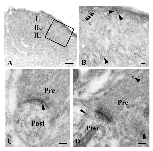
Figure 3.
Localization and distribution of PICK1 immunoreactivity in the spinal cord. A, PICK1 immunoreactivity in the dorsal horn was strongest in lamina I and inner lamina II (IIi). IIo: outer lamina II. B, High magnification of the outlined region from A. PICK1-positive somata (arrows), punctation, and patches (arrow heads) were observed. C, Immunogold labeling for PICK1 is prominent in the postsynaptic density (arrow). D, Immunogold labeling for PICK1 is prominent in the presynaptic terminal (arrows) and postsynaptic dendrite (arrowhead). Pre: presynaptic. Post: postsynaptic. Scale bars: 100 μm in A, 5 μm in B, 100 nm in C and D.

