Fig. 1.
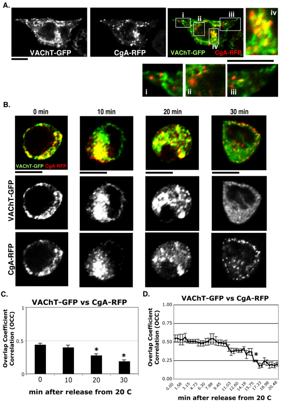
VAChT–GFP and CgA–RFP pass through a common intermediate compartment. (A) PC12 cells transfected with VAChT–GFP (green) and CgA–RFP (red) under steady state conditions at 37°C were imaged. The magnified insets i–iii and iv show VAChT–GFP and CgA–RFP at the cell periphery and at the cell center, respectively. (B) PC12 cells transfected with VAChT–GFP (green) and CgA–RFP (red) were incubated at 20°C to arrest Golgi-to-PM trafficking and then moved to 37°C to induce it. (C) Overlap coefficient correlations (OCCs) between VAChT–GFP and CgA–RFP were measured at 0, 10, 20, 30 minutes after 20°C block and release. The average OCC ± s.e.m. was calculated from three different experiments (n=30). (D) The real-time OCCs between VAChT–GFP and CgA–RFP for 20 minutes after 20°C block and release were measured (n=6 cells). Average OCC ± s.e.m. at every fifth shot from the beginning to the end of the time-lapse movie (supplementary material Movie 1) was picked and shown on the line graph (*P<0.05). Scale bars: 5 μm.

