Fig. 4.
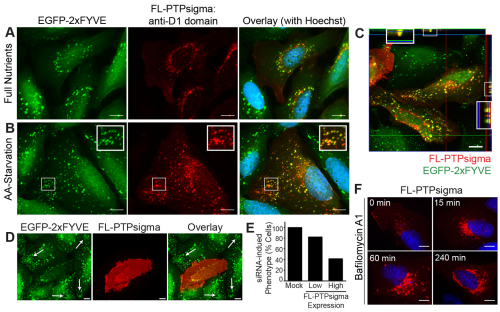
Exogenous PTPσ localizes to PtdIns3P vesicles and rescues the siRNA phenotype. (A,B) FL-PTPσ was transiently expressed in U2OS EGFP–2xFYVE cells and PtdIns3P and PTPσ imaged by fluorescent microscopy following incubation for 2 hours with full nutrient medium (A) or amino acid starvation medium (B) [green, PtdIns3P; red, anti-PTPσ (D1-targeted antibodies); blue, nuclei]. Insets are 2× magnifications of boxed regions. Scale bars: 10 μm. (C) U2OS EGFP–2xFYVE cells transfected with FL-PTPσ and amino acid starved for 2 hours were imaged using D1-targeted PTPσ antibodies. A Z-stack of 0.25 μm increments was captured using sequential channel acquisition and confocal microscopy, with the third slice displayed and Z-stacks through the X and Y planes shown at the border. Insets are 2× magnifications of boxed regions. Scale bar: 10 μm (green, PtdIns3P, EGFP–2xFYVE; red, anti-PTPσ; yellow, colocalization). (D,E) U2OS EGFP–2xFYVE cells were transfected with PTPRS siRNA-1 for 48 hours, after which FL-PTPσ (which lacks the sequence targeted by siRNA-1) was introduced for an additional 24 hours. PtdIns3P and PTPσ were imaged as previously described. The presence of siRNA-induced phenotype (abundant, non-perinuclear EGFP–2xFYVE-positive vesicles; indicated by white arrows in D) was determined for cells expressing no, low or high levels of FL-PTPσ (E). Scale bars: 10 μm. (F) FL-PTPσ-positive vesicular structures accumulate when lysosomal fusion is inhibited. U2OS cells expressing FL-PTPσ for 24 hours were treated with 100 nM Baf-A1 in full nutrient medium for 0, 15, 60 or 240 minutes and FL-PTPσ imaged using D1-targeted PTPσ antibodies (red). Nuclei were stained with Hoechst 33342 (blue). Scale bars: 10 μm.

