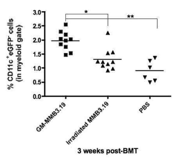
Figure 4.
Percentage of CD11c+eGFP− cells in the skin-draining LN of vaccinated mice. B6 mice were vaccinated with a single dose of 2×10⁵ GM-MMB3.19, non-GM-MMB3.19 cells or PBS alone. One week later, mice were exposed to 8.5 Gy and 4 h later transplanted with ATBM and lymphocytes from eGFP+B6 syngeneic mice in order to be able to differentiated between host (eGFP−) and donor cells (eGFP+). Three weeks post-BMT, cell suspensions from the skin-draining LN of individual mice were prepared for flow cytometric analysis to determine the percentage of CD11c+ expressing cells. Data were pooled from 2 separate experiments consisting of 3-6 mice per group. Statistical difference between groups was determined using Kruskal-Wallis one-way ANOVA analysis (p < 0.01) followed by Dunn’s multiple comparison test on individual pairs (*p < 0.05, ** p < 0.01).

