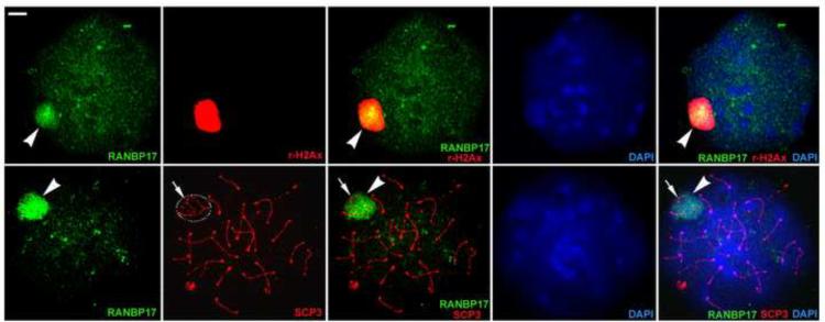
FIG. 4.
Immunofluroscent detection of RANBP17 in chromosome surface spreads of pachytene spermatocytes. In pachytene spermatocyte chromosome spreads, RANBP17 immunoreactivity (green and arrowheads) is predominantly concentrated in the XY body, which is strongly labeled by γH2Ax immunoreactivity (red in upper panels). Synaptonemal complexes of meiotic chromosomes are labeled as red fluorescence by the anti-SCP3 antibody and the XY body represents a sub-nuclear domain that can readily be distinguished based upon the shape of the SCP3-positive structures of the sex chromosomes (circle and arrows in lower panels). RANBP17 immunoreactivity is predominantly localized to the XY body. All panels are in the same magnification. Scale bar = 1μm.

