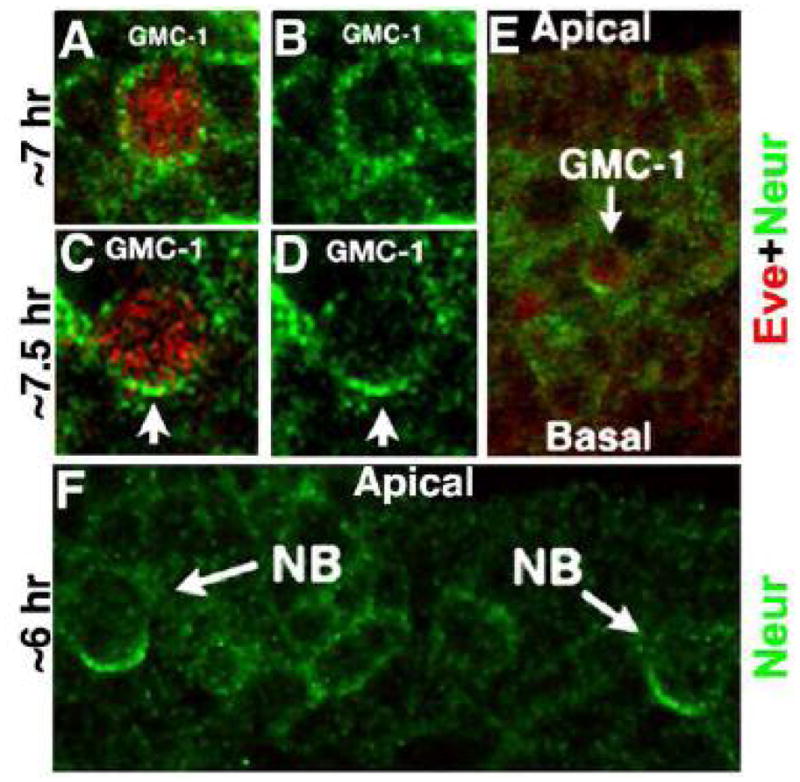
Figure 2. Neur is asymmetrically localized to the basal end in a GMC-1 prior to its division.

Embryos are double stained with Eve (Red) and Neur (Green) antibodies. Panels A–D are of the same magnification and a late GMC-1 (panels C, D) is larger than a mid GMC-1 (see Gaziova and Bhat, 2009). As shown in panels A and B, while Neur is less asymmetric and more uniform in a mid-stage GMC (a GMC-1 is normally born around 6–6.5 hrs of age), it is asymmetrically localized to the basal end of a late GMC-1 (panels C,D and E). Several NBs in a hemisegment also show a basally localized Neur (panel F), however, NB4-2 has no Neur expression. The GMC-1 development (timing) can be distinguished as an early, mid and late GMC-1 by looking at the development of the aCC/pCC lineage, appearance of cells of the EL lineage, and the migratory position of a GMC-1 within the nerve cord and the levels of expression of such proteins as Pdm1 and Pdm2. Moreover, a late GMC-1 is larger than a mid or an early GMC-1, and a mid GMC-1 is smaller than an early GMC-1 (see Gaziova and Bhat, 2009; see also Fig. 5).
