
Figure 5. Over-expression of Neur in a GMC-1 down-regulates the levels of Pdm1.
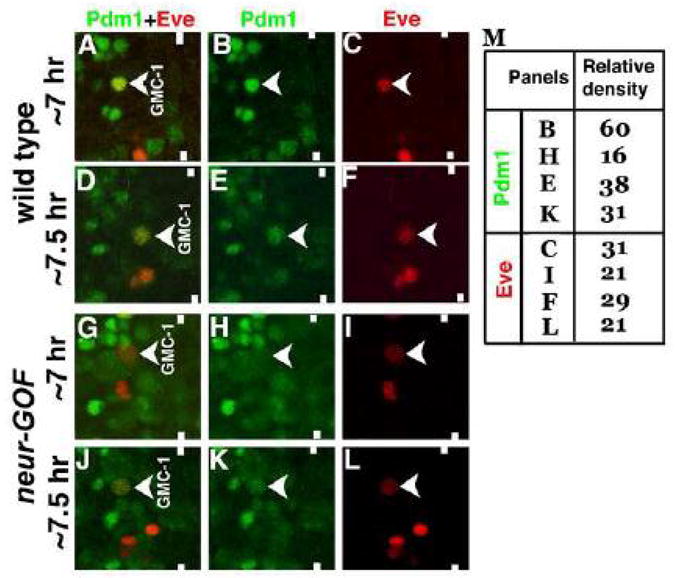
Embryos are double stained with Eve (Red) and Pdm1 (Green). Anterior is up, midline is marked by vertical lines. Pdm1 is expressed at high levels in a GMC-1 from a 7 hr old embryo (Panels A and B). The level drops as the development proceeds (Panels D and E). In neur-GOF embryos, with the induction of neur prior to the formation of GMC-1, the level of Pdm1 is drastically reduced as seen in a GMC-1 from a 7 hr old embryo (Panels G and H). The level of Pdm1 improves in a GMC-1 from a 7.5 hr old embryo (Panels J and K). In panel M, quantification of Pdm1 and Eve in GMC-1 was done by measuring the relative densities of Pdm1 and Eve staining signals in GMC-1.