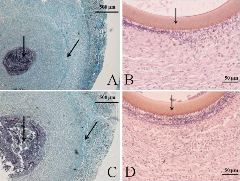
Figure 3.
Representative histology slides of cross sections stained with Masson's trichrome (A and C) or hematoxylin and eosin (B and D) of NO-releasing (A and B) and control (C and D) probes explanted at 14 d. Arrows in the hematoxylin and eosin stained pictures indicate the probe membrane. Arrows in the Masson's trichrome stained pictures indicate the implant site, surrounded by dark stained inflammatory cells and the collagen capsule. An increased capsule size and inflammatory response at the membrane surface are observed at control probes.

