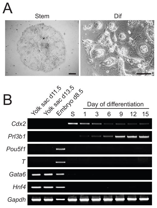
Fig. 1. Derivation and characterization of stem cell populations from rat blastocysts.
A) A colony of tightly packed cells characteristic of the stem cell state (Stem) and a colony of cells possessing larger nuclei following 8-days of differentiation (Dif). Differentiation was induced by culturing cells in the absence of FGF4, heparin, and REF conditioned medium. Scale bars, 250 μm. B) Germ layer lineage analysis. RT-PCR analysis for lineage markers in the rat blastocyst-derived stem cells in the stem cell state (S) and following 15 days of differentiation. Lineage markers: Cdx2 (trophoblast - stem cell), Prl3b1 (trophoblast - differentiated), T (mesoderm), Pou5f1 (epiblast), Gata6 (endoderm), and Hnf4 (endoderm). RNA isolated from yolk sac and embryos were used as controls for the extraembryonic endoderm and embryonic gene markers, respectively. Gapdh was used as an internal control.

