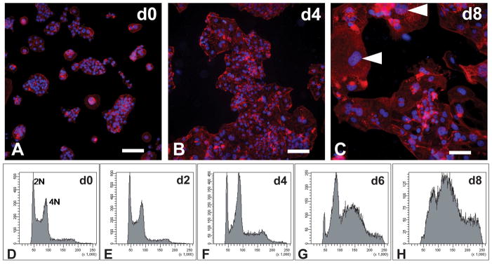
Fig. 3. Rat TS cell differentiation: morphology and endoreduplication.
Rat TS cells were cultured under stem cell conditions (d0; A, D) and differentiation conditions for 2 days (E), 4 days (B, F), 6 days (G), and 8 days (C, H). A–C) The cells were fixed, stained with rhodamine phalloidin, and counterstained with DAPI. Arrowheads show the presence of differentiated cells with trophoblast giant cell morphologies. D–H) DNA contents of the cells were determined by flow cytometry following propidium iodide staining. Scale bar, 100 μm.

