
Figure 2.
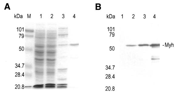
Expression and purification of Myh protein. The 10% SDS–polyacrylamide gel contains the following samples: lysate of CC104mutY/pREP4 cells containing pQE30 (lane 1) or pQE30/Myh (lane 2) after induction of Myh protein expression by IPTG; fraction eluted from an SP Sepharose column in the presence of 0.5 M KCl (lane 3); Myh protein eluted from a Ni2+–NTA column in the presence of 0.2 M imidazole (lane 4). The gel was visualized by Coomassie blue staining (A) and by western blot using polyclonal anti-Myh antibody (B). Myh protein is indicated on the right. Lane M contains molecular mass standards (Bio-Rad) as indicated on the left.