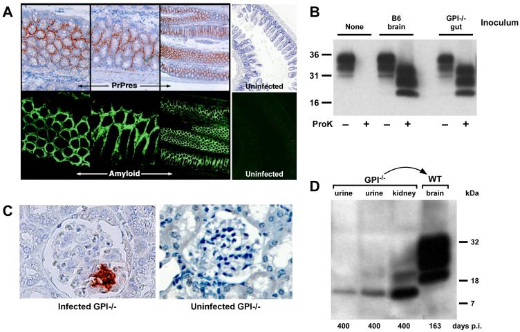
Fig. 2.
(A) PrPsc (PrPres) staining in the gut by immunohistochemistry. Sections of gut tissue from GPI−/− PrP tg mice were stained for PrPsc and amyloid by D18 and Thioflavin S staining, respectively (50x magnification). Note that PrPsc appears to deposit exclusively as amyloid in the gut. Uninfected control stains are shown on the right (50x magnification). (B) Western blot of brain homogenates of tga20 mice infected with either infected B6 brain homogenate or infected GPI−/− PrP tg mouse gut homogenate. Proteinase K digestion confirms that the PrP in the tga20 brain is PrPsc. (C) Immunohistochemical staining of the kidney reveals PrPsc deposition in the glomerulus after 400 days of infection (400x magnification). An uninfected GPI−/− PrP tg mouse kidney is shown as a control (400x magnification). (D) Urine and kidney tissue were taken 400 days post-infection and was transferred into B6 mice by intracranial inoculation. Both the urine and kidney samples contained PrPsc, as shown by Western blot, and were able to pass infection into a B6 mouse, as shown by the development of a proteinase K-resistant band pattern of PrP in the brain tissue of the mouse. The B6 mice also succumbed to disease after 163 days of infection, fitting with previous observations of PrP infection in B6 mice.

