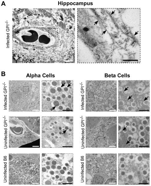
Fig. 6.
Electron microscopy of PrP in brain and islet cells of the pancreas. (A) PrP stained with D18 monoclonal antibody coupled to immunogold is visible in the hippocampus of GPI−/− mice infected with RML scrapie i.c. The right panel is a magnification of the region outlined with a dotted square on the left. Arrows point to PrP-labeled amyloid fibrils. (B) PrP is seen predominantly in the alpha cells of the pancreas in infected GPI−/− mice with deposition also noted in the beta cells (top panels). Little or no deposition is noted in uninfected mice, either GPI−/− (middle panels) or wild-type B6 (lower panels). The panels to the right are magnifications of the region outlined with a dotted square in each figure. Arrows point to gold-labeled PrP in the cells of the islets. The white scale bars are 2 um, while the black scale bars are 500 nm.

