
Fig. 6.
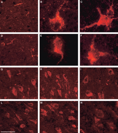
VGF peptide immunolocalisation in the bovine brain. VGF immunoreactivity (Cy3 red labelling) in frontal (A,D,G,L), parietal (B,E,H,M) and temporal (C,F,I,N) cortices. C-terminus immunoreactivity found in axons of the external layer (A) and in large perikarya with visible apical dendrites (B,C). NERP-1 antiserum labelling axons of the external layer (D), and perikarya with basal (E) and apical (F) dendrites. N-terminus (G–I) and TPGH (L–N) antisera-labelled groups of perikarya with different sizes and morphologies. Scale bar: 50 μm.