
Fig. 7.
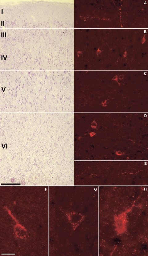
VGF peptide immunolocalisation in the rat brain. Immunoreactivity and cortical distribution of the C-terminus and NERP-1 peptides in the rat parietal cortex (Cy3 red labelling). (A–E) C-terminus staining. Roman numerals indicate cortical layers in cresyl violet sections (on the left), in parallel with immunofluorescence-stained sections. In all of the layers, the antiserum decorated either axons as perikarya found singly or in small groups, often with apical dendrites and sometimes disposed in a horizontal plane (B). Scale bars: 100 μm. (F–H) NERP-1 labelling was restricted to single perikarya with a granular staining and often with visible apical dendrites (F,H). Scale bar: 25 μm.