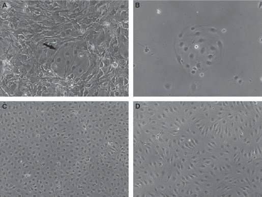
Fig. 1.

Phase contrast microscopy of human dermal microvascular endothelial cells (HDMECs). (A) Prior to purification, contaminants constitute the majority of the obtained cell population. Arrow indicates a group of HDMECs. (B) An isolated group of HDMECs after purification with CD31. The cells have a polygonal shape. Typical cobblestone morphology at confluence of blood (C) and lymphatic (D) HDMECs after separation of lymphatic HDMECs from the bulk of CD31+ cells with D2-40. Original magnifications: A and B, ×20; C and D, ×10.
