
Fig. 4.
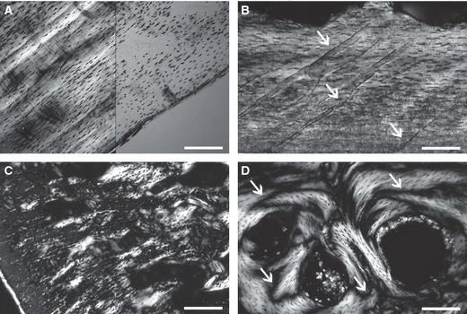
Longitudinal sections of (A, C, D) Varanus exanthematicus and (B) Varanus griseus. (A) Primary (pseudolamellar) periosteal bone in polarised (left) and natural (right) light. Note the mass birefringence and the parallel spindle-like osteocytes. Scale bar = 0.3 mm. (B) Primary periosteal bone in polarised light. The arrows point to vascular canals. Howship's lacunae are visible on the top of the primary formation. Scale bar = 0.2 mm. (C) Calcified cartilage in the core of trabeculae of endosteo-enchondral origin in polarised light. Scale bar = 0.3 mm. (D) Highly remodelled lamellar bone in polarised light. The arrows point to cementing lines of resorption. Scale bar = 0.2 mm.