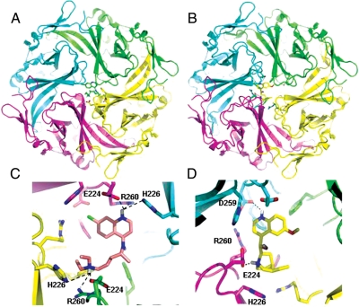
Figure 1.
Models of the lowest energy binding complexes. (A) Kir2.1 complexed with chloroquine. Kir2.1; ribbon structure with each chain depicted in a different colour. Chloroquine; green sticks. (B). Kir2.1:quinidine complex. Quinidine: yellow sticks. (C) Detailed view of chloroquine binding with Kir2.1 side chains within 5 Å of chloroquine (sticks.) (D). Close up view. Binding site of quinidine with side chains of Kir2.1 residues within 4 Å of the molecule (sticks). Hydrogen bonds: dashed lines. Kir2.1 amino acids are labelled as D259; F254; E224; R260, and H226.

