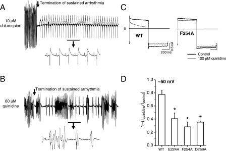
Figure 5.
Arrhythmia termination by 10 μM chloroquine and 60 μM quinidine. (A and B). 10 s ECG runs highlighting the termination of burst pacing-induced tachyarrhythmia by chloroquine and quinidine. The underlined regions are magnified beneath. Choroquine restored sinus rhythm. Quinidine was proarrhythmic. Alanine scanning mutagenesis of amino acids facing the cytoplasmic pore of Kir2.1. (C) Voltage command: 500 ms steps from −80 mV to −50 mV then to −100 mV, in the presence or absence of 100 μM Quinidine. Representative currents elicited in HEK cells expressing WT Kir2.1 (left), Kir2.1 F254A (right). Scales: 200 ms, 0.5 nA. (D) Fractional blocked outward current measured at −50 mV in the presence of E224A, F254A, and D259A. *<0.05, n= 5 cells each.

