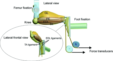
Figure 1. Schematic diagram of the rat hindlimb muscles and experimental situation for testing overload effects on contractile characteristics of m. tibialis anterior (TA) and m. extensor digitorum longus (EDL).

The lateral view shows hindlimb muscles after removal of the m. biceps femoris. Abbreviations: GL, m. gastrocnemius lateralis; TA, m. tibialis anterior; PE, peroneal muscles; EDL, m. extensor digitorum longus. Force–length characteristics of the TA and EDL were determined at 90 deg flexion of knee and foot. While the proximal insertions were left intact, tendons of TA and EDL were distally attached to force transducers via Kevlar® threads and aligned with their lines of pull in vivo. EDL was overloaded by (1) extirpation of TA, (2) tenotomy of TA and (3) TA ligament transection. Note that the retinaculum covering the tendons of the EDL and TA consists of two separate ligaments.
