
Fig. 10.
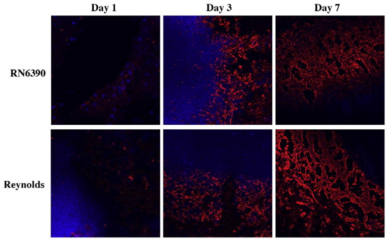
Fibronectin deposition along the developing brain abscess wall become prominent at day 7. Mice (n=4–6 mice per group/time point) were injected with either S. aureus strain RN6390- or S. aureus strain Reynolds- encapsulated agarose beads (104CFU) as described in the Materials and methods. Animals were euthanized at the indicated time points, whereupon brain tissues were flash frozen on dry ice for subsequent cryostat sectioning. Serial 10 μm thick sections were prepared throughout the entire abscess, subjected to immunofluorescence staining for fibronectin (red), and imaged using a 40× oil immersion objective lens by confocal microscopy. Nuclei were visualized using Hoechst 33342 (blue). Fibronectin immunoreactivity is shown along the peri-abscess area in the vicinity of the developing wall, where the dark areas represent necrotic regions. Results are representative of two independent experiments.