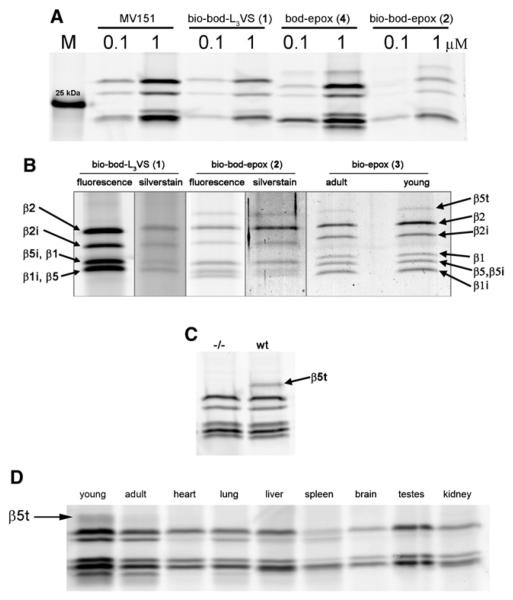
Figure 2. Activity-Based Profiling, Affinity Purification, and LC-MS Identification of Proteasome β Subunits in Murine Tissues Lysates.
(A) In-gel fluorescence detection of active proteasome β subunits in 3-week-old wild-type murine thymus homogenate after labeling with MV151, ABP 1, 2, and 4 (see also Figure S1). M indicates the molecular marker band of 25 kDa.
(B) In-gel fluorescence and silver stain detection of active proteasome β subunits in young and adult thymus after labeling with ABP 1, 2, 3, and affinity purification. Protein identification by LC-MS analysis of in-gel digested silver-stained bands (see Tables S1 and S2 for details).
(C) In-gel fluorescence detection with ABP 4 of β5t activity in wild-type and absence of activity in the (−/−) β5t knockdown thymus from 3-weeks-old mice.
(D) Activity-based proteasome profiling using ABP 4 shows β5t activity in murine thymus (young and adult) but not in heart, lung, liver, spleen, brain, testes, or kidney.

