
Figure 4.
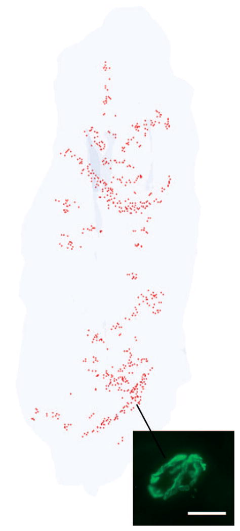
Three-dimensional reconstruction of all the α-bungarotoxin–positive NMJs in every 20th section through the entire thickness of a single control tibialis anterior muscle from an adult rabbit. All the NMJs are indicated in red, and the specimen is oriented with the origin and insertion at the top and bottom of the reconstruction. Inset is an example of an α-bungarotoxin–labeled NMJ. Total length of rabbit tibialis muscle is approximately 5.5 cm.