
Figure 7.
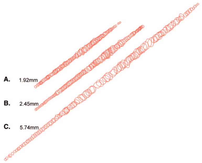
Representative examples of individual myofibers reconstructed from the (A) orbital layer, (B) global layer directly contiguous with the orbital layer, and (C) middle of the global layer. These represent the complete lengths of these three fibers followed in serial sections.