
Figure 9.
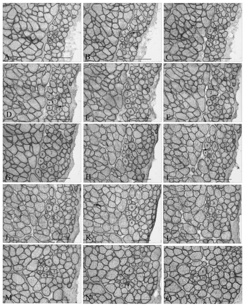
(A–O) Representative photomicrographs of a single fiber from an adult normal superior rectus muscle (4.622 mm long) from rabbit, demonstrating changes in its fascicular relationship with its neighboring fibers relatively often in the course of its fiber length. *Individual myofiber that was reconstructed. Numbered fibers allow observation of the changes in the neighboring muscle fibers over the course of 2.9 mm represented by these selected sections. Horizontal arrows indicate fibers that newly appear in the cross-sections within 2.9 mm of the reconstruction (numbers 9 and 10). Vertical arrow indicates a fiber that ends within 2.9 mm.