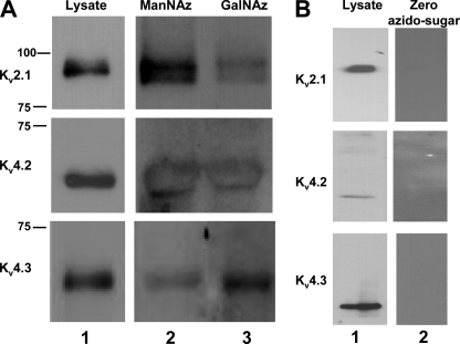
FIGURE 7.
Click-iT chemistry confirmed the presence of sialylated O-glycans attached to Kv2.1, Kv4.2, and Kv4.3. A, immunoblots of Kv2.1, Kv4.2, and Kv4.3 Pro5 lysates. Lane 1, untreated whole cell lysates; lane 2, Ac4ManNAz-labeled (sialic acid) samples; lane 3, Ac4GalNAz-labeled (O-glycosylated) samples. Molecular weight markers are noted to the left of lane 1. B, control treatment in the absence of azido-modified sugars. Lane 1, untreated whole cell lysates. Lane 2, biotin-treated, streptavidin-precipitated samples. Top, Kv2.1. Middle, Kv4.2. Bottom, Kv4.3.

