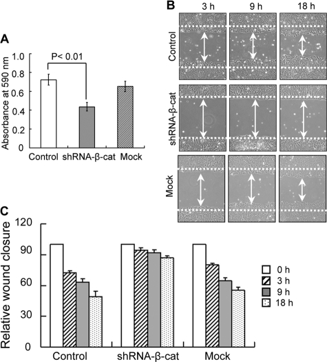
FIGURE 4.
Effects of β-catenin knockdown on fibronectin-mediated cell adhesion and migration. A, subconfluent cells were detached, and 40,000 cells were added to 96-well plates coated with 3 μg/ml fibronectin for the cell adhesion assay. The plates were incubated at 37 °C for 30 min and then washed twice with warmed PBS to remove non-adherent cells. The adherent cells were fixed with 25% glutaraldehydes and stained with 0.5% crystal violet, and then the absorbance at 590 nm was measured. Error bars, S.D. B, a confluent layer of cells was scraped/wounded using a yellow tip. The open gap was then inspected microscopically, and the distances between the sides of the wound were measured at the indicated times. C, quantitative data (mean value) for the cell migration from three independent experiments. The distance between the sides of the wound immediately following wounding (0 h) was set equal to 100. The relative wound closure is expressed as widths of the wound relative to the distance at 0 h.

