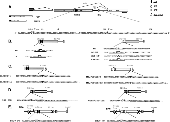
FIGURE 1.
PLP constructs. A, schematic of the PLP-neo splicing minigene construct. The arrows indicate the position of the PCR primers. The PLP and DM20 PCR products and partial sequences of PLP exon 3B (uppercase letters) and intron 3 (lowercase letters) in PLP-neo (WT) are shown; DM20 and PLP 5′ splice site are enlarged and in bold type; and G1, M2, and ISE are underlined. The filled ovals represent the wild type G1M2, empty ovals represent the G1M2 mutated to polyT, the shaded rectangle is the ISE, and the empty triangle represents the G runs of the ISE. B, schematics and sequences of the constructs in which the DM20 5′ss has been mutated to a stronger 5′ss: AU (canonical), GLO (α-globin), and C → A (disease-associated mutation (28). The sequences of natural DM20 5′ss and the strong 5′ss are underlined. C, schematics and sequences of the constructs in which the G runs of the ISE (empty triangle) are moved 6 nucleotides (PLPISE+6) and 12 nucleotides (PLPISE+12) downstream of the PLP 5′ss. The sequence of the ISE is underlined. D, schematics and sequences of the constructs in which the ISE replaces the M2. The G1, G1MT, and ISE sequences are underlined. E, schematics and sequences of the PY7 constructs generated for in vitro splicing and spliceosomal assembly (see “Experimental Procedures” for details). PLP exon 3A last 10 nucleotides, exon 3B, and intron 3 are cloned into the tropomyosin (Tropo) gene. The × indicates that the 5′ss of tropomyosin exon 2 and PLP 5′ss are mutated. DM20 WT contains the natural exon 3B sequences, whereas DM20 MT contains G1M2 mutated to poly Ts. SP6 refers to the promoter that drives in vitro transcription.

