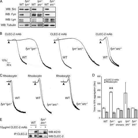
FIGURE 4.
A, whole cell lysates prepared from deficient platelets and their litter-matched wild-type platelets were immunoblotted with anti-Lyn, anti-Fyn, anti-Src, and anti-tubulin antibodies. B and C, washed platelets deficient in Fyn/Lyn, Fyn/Src, and Lyn/Src (2 × 10⁸ platelets/ml) were stimulated with 10 μg/ml CLEC-2 mAb (B) or 30 nm rhodocytin (C). Platelet aggregation was measured as a change in light transmission, using a lumi-aggregometer. Representative aggregation traces from three independent experiments are shown. The addition of the agonist is indicated by an arrowhead. D, data represent the means of the time to get 50% of aggregation and standard error of three independent experiments. **, p < 0.005 (significant difference versus wild type, according to two-tailed Student's t test). E, Fyn/Src-deficient washed platelets and their litter-matched wild-type platelets (2 × 10⁸ platelets/ml) were stimulated with 10 μg/ml CLEC-2 mAb for 3 min. CLEC-2 was immunoprecipitated (IP), and immunoprecipitates were immunoblotted with an anti-phosphotyrosine antibody. CLEC-2 immunoprecipitates were also immunoblotted with anti-CLEC-2 antibody as described under “Experimental Procedures.” Representative data from two independent experiments are shown. WB, Western blotting.

