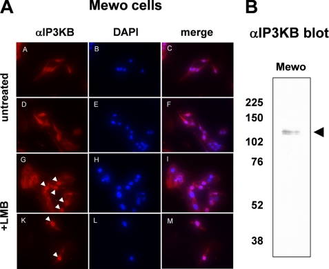
FIGURE 1.
Endogenous expression of IP3KB in mammalian cells. A, Mewo cells were either treated with 20 nm LMB for 6 h or mock treated. Then, intracellular localization of endogenous IP3KB was determined by immunofluorescence techniques using antibody H00003707-M01 (panels A, D, G, and K). Nuclei were stained with DAPI (panels B, E, H, and L), and overlays of the digitized images were created (panels C, F, I, and M). Nuclei showing a strong accumulation of IP3KB are marked by white arrowheads. B, cell lysates from Mewo cells were separated on SDS-polyacrylamide gels. Antibody H00003707-M01 was used to detect IP3KB using Western blot analysis. The black arrowhead indicates the expected protein band of endogenous IP3KB. The molecular mass of protein markers is indicated on the left in kDa.

