FIGURE 2.
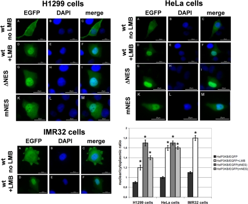
Exportin 1-dependent nuclear export of IP3KB. Different EGFP fusion genes were transiently expressed in H1299, HeLa, and IMR32 cells. After 24 h of expression, cells were fixed and nuclei stained with DAPI. EGFP fusion proteins (panels A, D, G, and K) and DAPI (panels B, E, H, and L) were visualized by fluorescence microscopy, and an overlay of the digitized images was created (panels C, F, I, and M). Cells expressing EGFP fusion proteins of full-length HsIP3KB (WT) were either untreated (no LMB) or treated for 6 h with 20 nm LMB (+LMB). In additional experiments, mutated forms of HsIP3KB lacking the NES (ΔNES) or possessing a functionally inactive NES (mNES) were expressed. In three independent experiments, at least 100 cells were analyzed for nuclear/cytoplasmic ratio of EGFP fluorescence (see “Experimental Procedures”). Results from one representative experiment are shown (mean ± S.E.) in the bar graph. For significance analysis, one-way analysis of variance and unpaired t test were used (*, p < 0.01; not significant, p > 0.05). Values were always compared with HsIP3KB/EGFP. For detailed information about the constructs, see Table 1.

