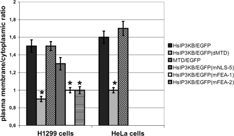
FIGURE 4.
Plasma membrane localization of IP3KB mediated by its binding to cortical actin. Different EGFP fusion genes of HsIP3KB were transiently transfected into H1299 and HeLa cells. Cells were fixed 24 h post transfection, and EGFP was visualized by fluorescence microscopy. WT, wild type HsIP3KB; MTD, isolated multi-targeting domain; ΔMTD, multitargeting domain deletion mutant; mFEA-1/2, FEA motif substitution mutants. The plasma membrane/cytoplasmic ratio was determined as described under “Experimental Procedures.” For detailed information about the constructs, see Table 1; for further details, see the legend to Fig. 2. Values were compared with HsIP3KB/EGFP.

