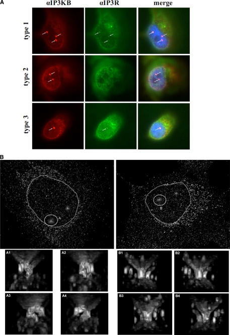
FIGURE 5.
Enrichment of IP3KB at subnuclear structures (A) H1299 cells were fixed, and endogenous IP3KB (red) was detected by immunofluorescence techniques using antibody H00003707-M01. In addition, Ins(1,4,5)P3 receptor type I, II, and III (green) was detected using isoform-specific antibodies. Nuclei were stained with DAPI (blue), and an overlay of the digitized images was created. Nuclear structures showing an accumulation of IP3KB are marked by white arrows. B, endogenous IP3KB in H1299 cells was detected by immunofluorescence techniques. Then, microscopic data sets were obtained by fluorescence microscopy and subsequent deconvolution. Three-dimensional reconstructions were created using the ImageJ plug-in three-dimensional viewer. Reconstructions of two exemplary H1299 cells are shown. Exemplary nuclear invaginations are marked by white circles (A and B), and magnified images from different perspectives (A1–A4 and B1–B4) are shown below. Nuclei are marked by dotted circles.

