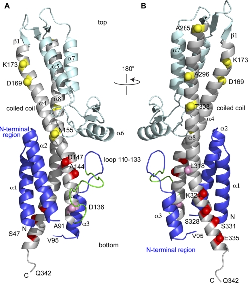
FIGURE 1.
Crystal structure of SipD (PDB ID 2NZZ) shown with the positions of the 14 spin labels (indicated as spheres) throughout the length of the central coiled-coil (helix α4 and α8). The spheres are colored based on the strength of the PRE effect with PrgI as yellow (weak), pink (moderate), and red (strong). The SipD N-terminal region (helix α1-α3) is colored blue, the central coiled-coil is colored gray, and the rest of SipD is colored cyan. A 16-residue loop in the N-terminal region (residues 118–133, colored green) lacked electron density and was modeled using CHARMM (33) and energy-minimized to remove steric clash. The SipD crystal starts at Ser-47 and ends in Gln-342, and residues 92–94 also lacked electron density. SipD in A is rotated by 180° in B.

